European foulbrood (EFB) is a statutory notifiable infection of honey bees. Any beekeeper in England or Wales who suspects the presence of AFB in a colony for which they are responsible is legally required to inform the NBU.
European foulbrood (EFB) is caused by the bacterium, Melissococcus plutonius. The bacteria are a gut pathogen and cause the death of some proportion of the larvae they infect. Some strains are very virulent, but other strains, like many of those found in the UK, are less virulent. However, lower virulence can lead to low-level or recurring infections.
This page describes some of the signs of EFB, for a more comprehensive understanding of EFB disease in honey bees, including how to recognise the signs of EFB and differentiate them from other brood diseases, please read our ‘foulbrood disease of honey bees’ advisory leaflet.
Signs of EFB include:
|
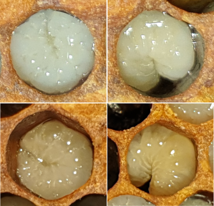
EFB infected larvae no longer sit in their cells in a healthy 'C' shape and lose their plump segmented appearance. Some become twisted in their cells and others have the appearance of looking melted down. |
|
|
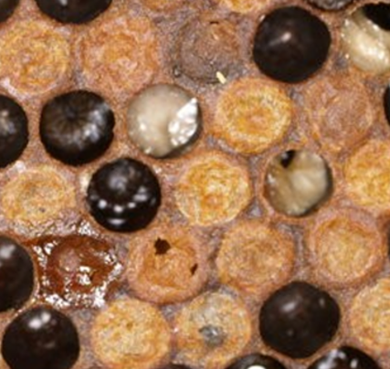
The gut of an infected larva may be visible through its translucent body wall. It has a creamy colour caused by the mass of bacteria living within it. This image shows some yellow looking larvae (black arrows) next to normal pearly-white larvae; these larvae have discoloured slightly and were not sitting neatly in their cells. |
|
|
Early on, the larval gut will begin to fill with bacteria. The black arrow in this image shows a small creamy mass in the larval gut; this is the bacterial mass. As the bacteria multiply, this mass expands and may take up most of the gut. |
|
After dying, the larval remains will turn brown. Sometimes infected larvae die after they are sealed. When this happens, there may be perforated cell cappings, where the nurse bees have detected the disease and investigate. |
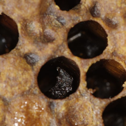
|
As dead larvae decompose further, they dry out and turn into a brown scale, which has a rubbery consistency, and can be easily removed from the cell. When there is a heavy infection with EFB, the brood pattern will become patchy and erratic as dead brood is removed by the nurse bees. |
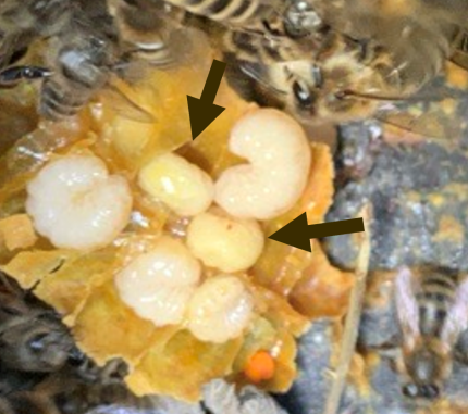 Image courtesy: Meg Seymour
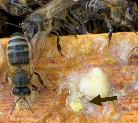 Image courtesy: Meg Seymour
To report a suspected case of EFB, please contact us as soon as possible at: [email protected]
Further information
Control of EFB is subject to the Bee Diseases and Pests Control (England) Order 2006: https://www.legislation.gov.uk/uksi/2006/342/contents/made
To learn more on the prevalence and distribution of EFB in England and Wales, view our disease incidence page: https://www.nationalbeeunit.com/diseases-and-pests/reports-charts-and-maps/disease-incidence/